Chapter 6: The Reticular Formation

Brain: Contents Page

The Reticular Formation                          Topics :     

The Reticular Formation

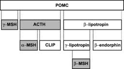
The brainstem contains many neurones that do not belong to well defined groups such as the cranial nerve nuclei or the olivary nuclei, or fibre tracts such as the pyramidal tract, the medial lemniscus or the medial longitudinal bundle. The reticular substance of the brainstem, the tissue between the distinct specific nuclei or tracts, appears to consist of a hotchpotch of nerve cells without obvious similarities, and axons that pass in many directions, intersecting with each other, rather than travelling together in the form of a tract.

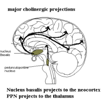
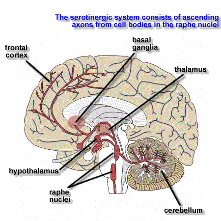
Modern histochemical techniques have shown that within the reticular formation there are groups of neurones with ill-defined boundaries that have characteristic chemical properties and neurotransmitters. Examples are the basal forebrain neurones that contain acetylcholine, and the raphe nuclei that contain serotonin. So there are subgroups of reticular neurones that contain specific neurotransmitters, and their axons have significant actions within the brainstem, forebrain and spinal cord.

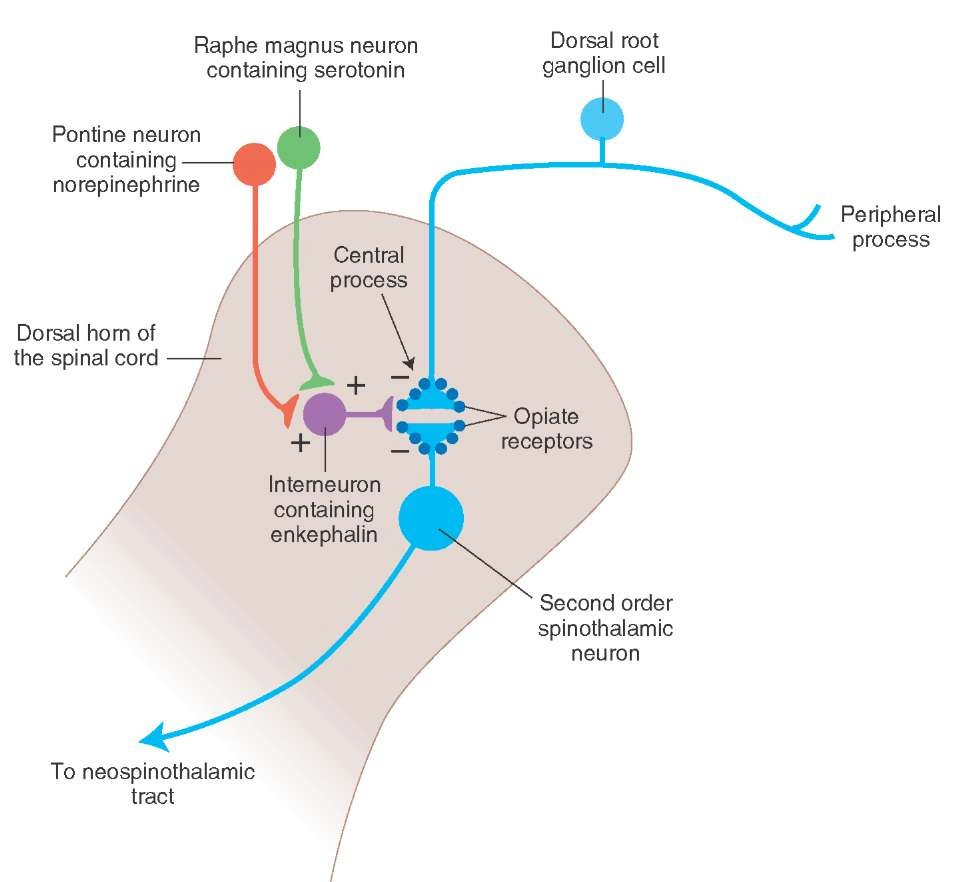
The reticular formation neurones receive inputs from many different systems - somatosensory, proprioceptive, auditory, visual, etc. These neurones have non-specific inputs, not concerned with signalling the precision found in the main sensory pathways, but they mix and integrate the activity of many systems to produce an overview of all on-going activity in the sensory systems. Some reticular neurones are concerned with regulating the activity of the forebrain, as in sleep, arousal and waking (this is known as the Ascending Reticular Activating System, ARAS). Others, the 'Descending Reticular Formation' modulate the activity of spinal circuits, in the regulation of muscle tone, autonomic outflow or transmission of nociceptive information in the dorsal horn.


Neurotransmitters used in the CNS, and Reticular Formation Pathways defined by their Neurotransmitters

Nerve cells use specific chemicals as neurotransmitters at their nerve endings.

Amino Acid Transmitters:

  • Glutamate is the most common excitatory neurotransmitter throughout the central nervous system. Aspartate is also an excitatory transmitter in many areas of the CNS.
  • Glycine is an inhibitory neurotransmitter in the spinal cord.

Within.the Reticular Formation there are groups of cells that project widely and these use Amines, Peptides or Acetylcholine as transmitters:

Aminergic neurones release one of the following amines at the terminals :

  • Dopamine, e.g. in is the Substantia Nigra.
  • Noradrenaline, e.g. the Locus Coeruleus
  • Adrenaline, e.g. some descending pathways from the reticular formation
  • 5-hydroxytryptamine (serotonin), e.g. pathways originating in the raphe nuclei in the midline of the brainstem
  • Histamine is the neurotransmitter in tuberomamillary neurones of the hypothalamus, which project to all parts of the CNS.
  • Gamma-amino-butyric acid (GABA) is the main inhibitory neurotransmitter in the CNS.

Some central neurones, e.g. the Basal Forebrain Nuclei, release acetylcholine, and these are assuming some importance as they degenerate in Alzheimer's Disease.

Peptides:

  • Substance P is present in small diameter afferent fibres in the dorsal horn.
  • Enkephalins are used in pathways concerned with pain control.
  • Orexin and Galanin are two others that have a role to play in the sleep-waking cycle.

Co-transmission using two transmitters, say, an amine and a peptide, has been shown in some pathways.

Many psychotropic and addictive drugs interact with these pathways.

*


Distribution of Neurotransmitter Pathways in the Central Nervous System   Top

Cholinergic Pathways

Nucleus basalis

Pathways that use acetylcholine within the brain include neurones in the basal forebrain concerned with arousal and the sleep/waking cycle including the Nucleus basalis, also called the Nucleus of Meynert. The nucleus basalis has projections that reach throughout the cerebral cortex.

This nucleus shows degenerative changes in Parkinson's Disease and Alzheimer's Disease, and acetylcholine production is reduced in some types of dementia. One focus for treatment of Alzheimer's disease is on ways to increase acetylcholine production.

Cholinergic Brainstem Nuclei

A second group of cholinergic neurones project to the thalamus from the brainstem (the pedunculopontine nucleus in the rostral pons).

Acetylcholine is also present in a number of brainstem nuclei that project to the caudate nucleus.

Cerebral Cortex

Within the cortex, cholinergic neurones also exist in the amygdala and hippocampus; the latter is of course associated with the laying down of memories.

*

Noradrenergic Pathways   Top

Noradrenaline (Norepinephrine) is released at the axon terminals of neurones of reticular formation nuclei such as the locus coeruleus (LC) and the lateral tegmental noradrenergic system.

Locus Coeruleus

Locus coeruleus neurones send their axons to the hypothalamus and throughout the cortex, including the amygdala, and the cerebellum.

They are concerned with the level of consciousness, stress and reward.

Brainstem

The noradrenergic brainstem nuclei send their axons to the spinal cord where they reach the dorsal horn, autonomic nuclei and the ventral horn. In addition the Nucleus tractus solitarius, involved in cardiovascular control, receives a noradrenergic projection.

*

Adrenaline (Epinephrine) is present in some neurones in the hypothalamus and brainstem concerned with stress and cardiovascular control.

All aminergic neurones synthesise their transmitters from the amino acid tyrosine.

Adrenaline is synthesised from noradrenaline (by methylation) and noradrenaline (also called norepinephrine) is synthesised from dopamine

It is produced as a result of methylation of noradrenaline, which , in turn, is a derivative of dopamine (by hydroxylation).

Although these amines are similar, they can have quite different actions in the central nervous system, and are released by separate neuronal pathways.

*

Dopaminergic Pathways   Top

Nigro-striatal Pathway

Dopamine is present in the midbrain and two dopaminergic pathways originate from the substantia nigra and the ventral tegmental area. The nigro-striatal pathway projects to the corpus striatum where it is concerned with the planning and control of movement.

Huntington's Disease, Ballism and HemiBallism, as well as Parkinson's disease are different types of movement disorder. A resting tremor is present in the hands of many patients with Parkinson's disease, and in Hemiballismus there are involuntary movements of the large joints, such as the whole arm.

Ventral Tegmental Pathways

There is also a significant domaminergic pathway that projects from the ventral tegmental area (VTA) to the frontal cortex, the hippocampus and the Nucleus accumbens of the basal forebrain, that has a role in mood, pleasure and reinforcement, and in addiction to drugs and alcohol.

Serotoninergic Pathways   Top

Serotonin (5-Hydroxytryptamine; 5-HT) is another amine with important actions on emotional behaviour. The cells of origin are particularly in the midline raphe nuclei which exist at different levels of the brainstem.

Rostral Raphe Nuclei

The axons of the rostral raphe nuclei are distributed throughout the cortex and in the hypothalamus and cerebellum. They are involved in the sleep-waking cycle.

Medullary Raphe Nuclei

The medullary raphe nuclei project to the spinal cord. Some of these descending axons terminate in the dorsal horn of the cord and have an anti-nociceptive influence on transmission in the dorsal horn.

This pathway ends on interneurones in the dorsal horn that synthesis and release enkephalin, a peptide neurotransimitter, and can alter the pain threshold of individuals (see below).

*

Histaminergic Pathways   Top

Histaminergic neurons in mammalian brain are located exclusively in the tuberomamillary nucleus of the posterior hypothalamus and send their axons all over the central nervous system.

Although in the hypothalamus, they are included here because of their their projection pattern, and because of their interaction iwth the other brainstem nuclei.

They are active only during waking and and induce and maintain wakefulness and attention, and the level of firing depends on the level of wakefulness/attention.

The ventrolateral preoptic area (VLPO) of the hypothalamus inhibits the activity of the histaminergic neurones and promotes sleep; this inhibitor pathways involves GABA..

*

Peptidergic Pathways   Top

There are many peptide neurotransmitters. Substance P is one, present in primary afferents concerned with nociception.

Others include Neuropeptide Y (NPY) and Cholecystokinin (CCK) and the Enkephalins (ENK).

In the hypothalamus, ENK are made as a product of the synthesis of AdrenoCorticoTrophic Hormone (ACTH) and Melanocyte Stimulating Hormone (MSH) from a peptide called Pro-Opiomelanocortin (POMC).

*

Beta-Endorphin can be released as a hormone, and is also the precursor of two enkephalins that are neurontransmitters. The illustration opposite shows the involvement of an ENK interneurone in the dorsal horn. Enkephalins act on opiate receptors, and this system is used in spinal anaesthesia, as local injection of low doses of opiates into the CSF helps manage pain.

*

Vasopressin and Oxytocin are hormones secreted by the neurohypophysisis.

Oxytocin is synthesised in the Paraventricular nucleus of the hypothalmus, and one part of this nucleus sends axons to the autonomic areas of the spinal cord, where oxytocin acts as a neurotransmitter rather than a hormone.

The pathways shown opposite indicate a role of the paraventricular nucleus and its spinal projection to the sympathetic, lateral horn of the thoracic cord. The pathways has significance in the control of the pineal gland and the secretion of melatonin, which follows the light-dark cycle. It is also involved in regulating the daily sleep/waking cycle.


*
HL Hass 2008 Physrev.Physiology.com
Wikipedia Commons
neurology.org
docstoc.com
what-when-how.com
www.unifr.ch
www.flyfishingdevon.co.uk
Wikipedia Commons
www.studentconsult.com
mikeclaffey.com

Top


Chapter 6: The Reticular Formation

Brain: Contents Page

The Reticular Formation                          Topics :     

© HumanPhysiology.Academy 2014-2015